顱后窩外科應用解剖
顱后窩外科應用解剖
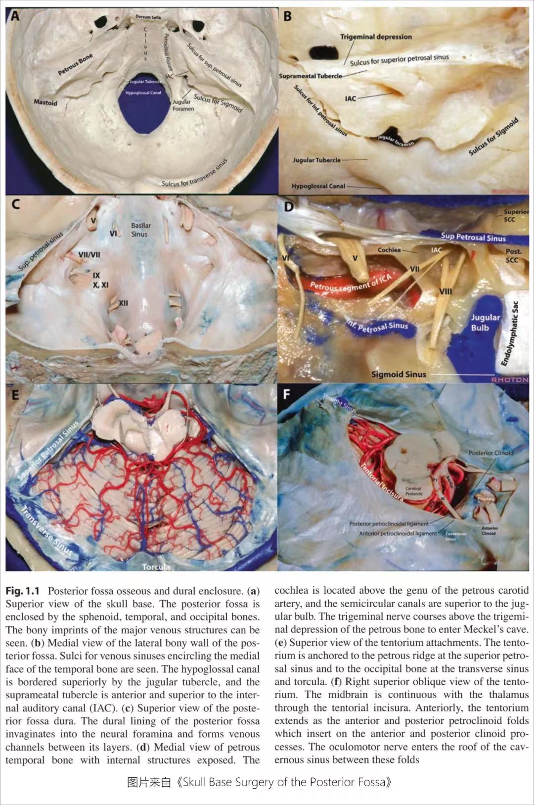
顱后窩的外科解剖(surgical anatomy of the posterior fossa)
顱后窩僅占顱腔容積的1/8;它通過小腦幕切跡(Tentorial hiatus)與幕上相通,通過枕骨大孔(foramen magnum)與椎管腔相通;其內容納小腦、腦干及大部分顱神經。
顱后窩的前、后、外、上等各個面同時兼具手術通道的作用,各個面的邊緣常存在硬腦膜靜脈竇,這些靜脈竇兼具界定顱后窩手術范圍的作用。眾所周知,橫竇及小腦幕以上的顱腔為幕上腔,可以理解為橫竇是顱后窩后部的上界,枕骨和頂骨之間的骨縫即人字縫走行在顱后窩邊界的上方,所有顱后窩的手術都應在橫竇以下的范圍內切開顱骨完成手術,即所謂的枕下開顱。
根據個人學習需要對以下相關外科解剖知識點進行復習:顱后窩的面、靜脈竇、靜脈竇在顱骨表面的定位、枕下顱骨及枕下肌群。
(一)顱后窩各個面的組成:
1、前:
(1)鞍背(dorsum sellae)
(2)蝶骨體的后部(posterior part of the sphenoidbody)
(3)枕骨的斜坡部(clival part of the occipital bone)
2、后:枕骨鱗部(squamosal part of the occipitalbone)的下份
3、兩側:
(1)顳骨的巖部(petrous parts of the temporalbone)
(2)顳骨的乳突部(mastoid parts of the temporalbone)
(3)枕骨的外側部(lateral part of the occipitalbone)
(4)后上方的小部分頂骨乳突角(mastoid angle of theparietal bone)
4、上:
(1)小腦幕(tentorium)
(2)小腦幕裂孔(Tentorial hiatus)=小腦幕切跡(tentorial incisura)
5、下:枕骨大孔(foramen magnum)


